

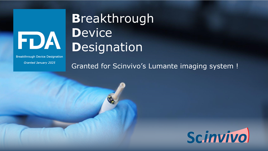
Breakthrough Device Designation
We’re very proud to share that Scinvivo’s Lumante Imaging system has received FDA Breakthrough Device Designation!
The Breakthrough Devices Program is a voluntary program for medical devices that provide for more effective treatment or diagnosis of life-threatening diseases or conditions, and its intention is to provide patients and health care providers with timely access to medical devices.
Scinvivo’s Lumante imaging platform is intended for realtime, minimally invasive visualization and local staging of bladder cancer.
Our mission
Worldwide, the incidence of cancer is growing, as life expectancy increases. Current imaging tools have their limitations as they are not suitable to provide the necessary information during diagnosis. Scinvivo aims to revolutionize cancer diagnostics and care by providing medical professionals with the next generation minimal invasive imaging platform. This imaging platform fills the gap that is left open by the current imaging technologies, as current technologies are unable to show the anatomical structure of the tissue during endoscopic procedures.
Scinvivo solves unmet needs for Payers, Patients and Doctors

Best possible care for patients, while costs are reduced

Improved quality of life by reducing invasive surgeries

Improved cancer diagnosis and treatment
Our solution
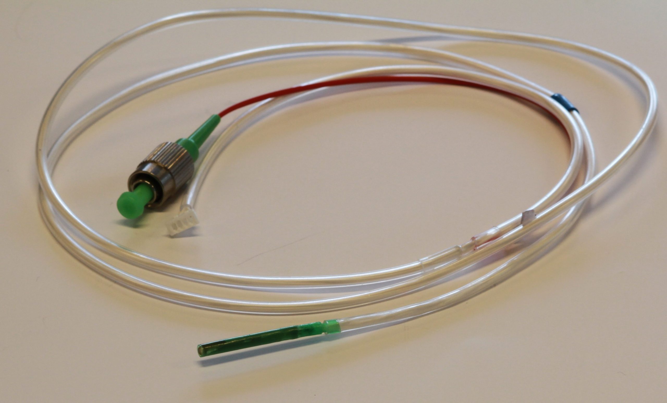
Scinvivo first focusses on the medical needs in bladder cancer. Scinvivo’s forward-looking OCT-catheter provides the answer by giving urologists real time ultra high resolution (a few micrometers) insight in the anatomical structure during diagnosis (cystoscopy).
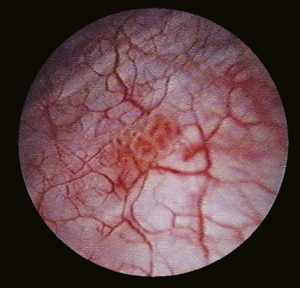
Classical cystoscopy image on which a tumor is visible
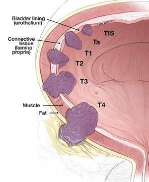
Different stages (TIS-T4) of bladder cancer
Disclaimer: Clinical investigations are pending to cover these claims. Scinvivo products do not have CE or FDA clearance yet.
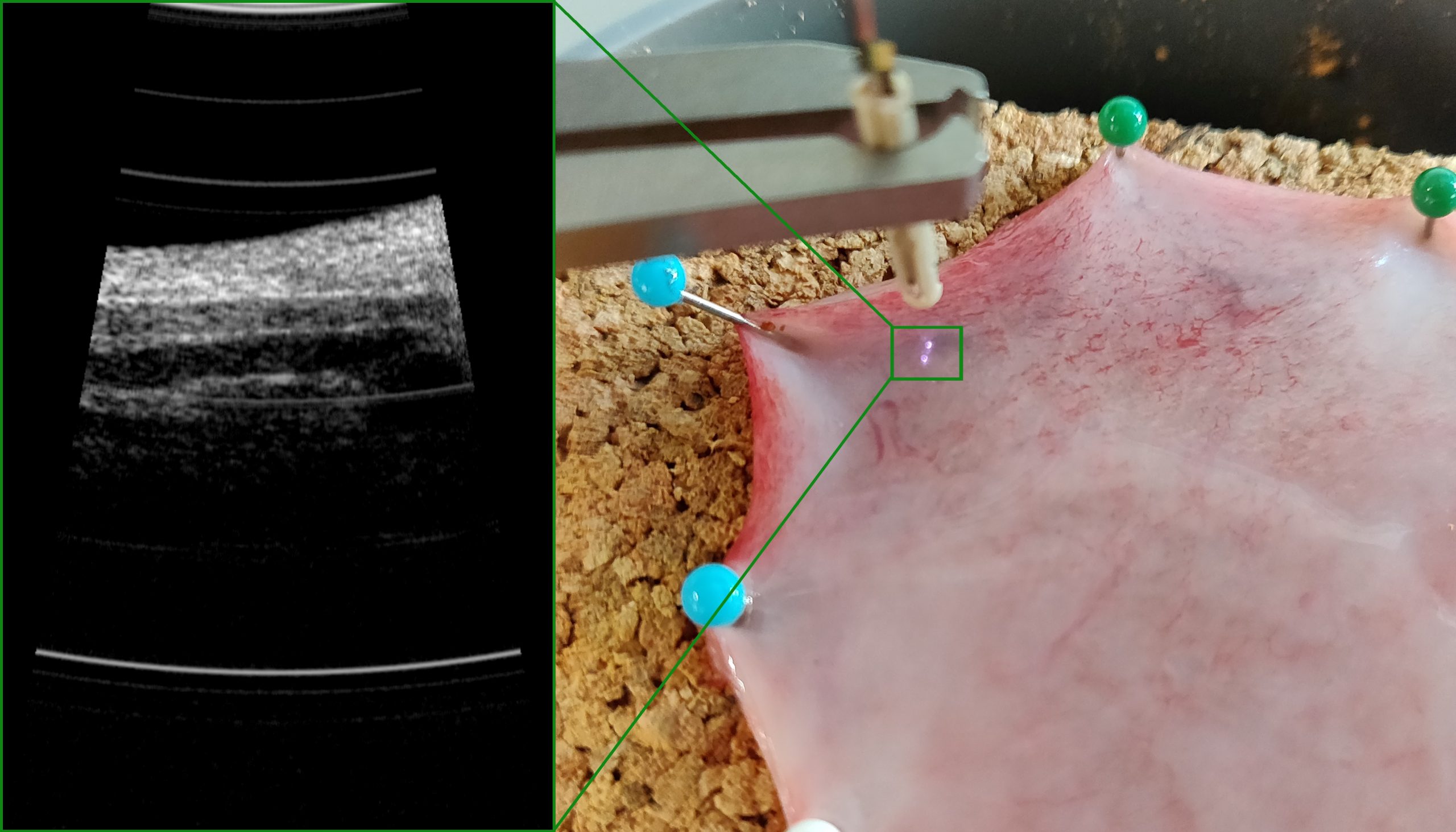
Our Product
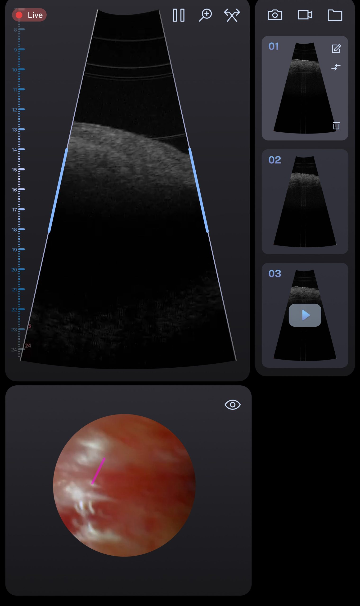
In the image below the first results of our second generation OCT probes is visible. An OCT image (left) made with one of our probes of porcine bladder tissue (right). The different layers of the bladder wall are clearly visible, as well as small blood vessels running through it.
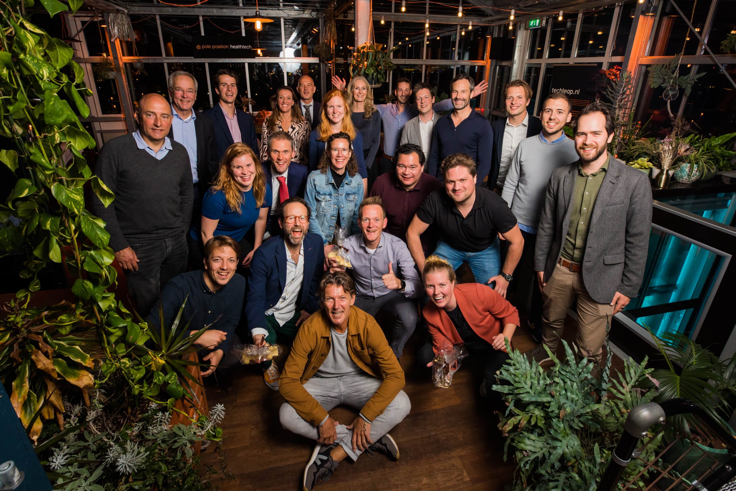
Our team
CMO
Maaike de Jong works as CMO (chief Medical officer) for Scinvivo since it was founded. As CMO, she is continuously developing the imaging platform and arranging the (pre)-clinical trials. Next to that, she secures funding for Scinvivo in the form of grants, loans, and new investments. Over the years Maaike gained experience in raising new capital and added a valuable contribution in the deal closing process. She gained significant project management experience by leading a Eurostars project, and an EFRO project. She holds a MSc in Biomedical Engineering and worked in Boston at Massachusetts General Hospital to specialize in Optical Coherence Tomography and its endoscopic applications. Maaike has an extensive international network regarding OCT technologies and urology. For the regulatory affairs, both CE & FDA, Maaike gained important experience and knowledge for Scinvivo and she is the main contact for the urologists in our network.
She has a strong belief that the imaging platform Scinvivo develops will be a game changer in cancer diagnostics.
CEO
Marijn van Os is CEO at Scinvivo, since October 2019 and co-founded of Scinvivo in 2016. As an entrepreneur he also founded Innoluce in 2010 as a spin-off of Royal Philips, including acquiring the main MEMS mirror IPR. Marijn successfully managed the acquisition of Innoluce by Infineon in 2016 for the automotive applications. Prior to this acquisition the medical assets, IPR and knowhow was transferred to Scinvivo. Marijn holds a PhD in mechanical engineering and continued his working career at Philips where he held different positions as technologist and development manager within The Netherlands and in the Czech Republic. Marijn has an extensive international network in the MEMS space and in the medical space for Urology.
Marijn is always looking for opportunities to improve and challenge the status quo and as such he is convinced that with the solution of Scinvivo the unmet needs in the cancer diagnostics & care will be improved significantly for all stakeholders, but in particular for the patients.
CTO
Camile van der Heijden joined Scinvivo in June 2018 and fulfils the role of CTO. Camile studied Biomedical Engineering (Bachelor) and Medical Engineering (Master). Camile holds a PDEng title Qualified Medical Engineer from the Stan Ackermans Institute. Prior to working for Scinvivo, Camile was employed at the Maxima Medical Center. There he worked on designing an improved educational simulation environment for use during training of the Neonatal Invasive Care Staff. Camile has a great network in the medical field in the Netherlands and is able to oversee the impact of product design on the medical regulatory application. While working for Scinvivo, Camile has learnt to manage suppliers through relationships built on mutual trust. In his role at Scinvivo he is responsible for product & process design, and works closely with our assembly partners to translate prototypes into repeatable products.
He believes technology has the potential to change lives for the better and it inspires and motivates him to help make that potential a reality.
CCO
Liesbeth Geerts joined Scinvivo October 2024 as CCO. Liesbeth holds a PhD in biomedical engineering from Eindhoven University of Technology and worked at Philips for over 22 years. At Philips, Liesbeth held various positions in the MRI business, from application specialist to clinical scientist and department manager. Through this wide experience, she gained a thorough understanding of the role of medical imaging in diagnosis and treatment and the way of working in hospitals. She has experienced how to inspire and steer innovation, and how to bring innovations to the end-user.
Liesbeth has collaborated with clinical partners across the globe, which has given her insight in region-specific clinical needs and preferences, and a strong clinical network.
It is Liesbeth’s ambition to have a positive impact in people’s lives by bringing innovations for better, faster and more patient friendly care to market. If done well, these innovations also benefit society by lowering overall healthcare cost. Liesbeth believes that Scinvivo’s imaging platform addresses both these needs, and is therefore excited to help bring this product to market.

CFO
Maarten Haast joined Scinvivo in February 2025. Maarten has a background in accountancy and holds en executive master of Finance & Control from the Vrije Universiteit in Amsterdam and worked at Stryker (US listed multinational in the Med Tech industry) over 20 years. Maarten held various positions from Financial Controller to CFO for the EEMEA division.
At Stryker Maarten has been heavily involved in many projects varying from implementations of different ERP systems, implementation of a European logistics center in Venlo, implementation of a COE and financial shared service center, acquisition and integration of different newly acquired companies.
Maarten has a strong preference to work in the Med Tech sector ultimately to help improving patients’ lives.
Next to his role as part-time CFO he is also active in advising SME entrepreneurs in M&A
Medical System Engineer
Maria Frías joined Scinvivo in November 2022 as Medical System Engineer. Her role is to support the system’s integration, validation and verification tests, documentation development for medical certification and support with supplier management. She holds a Master in Biomedical Engineering, and has 8 years of experience as application engineer and system engineer in the automotive and medical technologies fields. She has worked in different R&D companies where she gained experi-ence in European projects, system development, test development and integration, and managing suppliers and stakeholders.
She believes that Scinvivo’s technology provides medical professionals a tool to progress in cancer research, save more patients, and improve quality of lives.
QA/RA Manager and PRRC
Sonja Holten is the QA/RA Manager and PRRC for Scinvivo. At Scinvivo she is responsible for overseeing Quality Assurance and Regulatory Affairs as well as ensuring compliance with medical device regulations for all products and services. Sonja brings extensive experience in navigating the complex regulatory landscapes and implementing robust quality management systems. She plays a pivotal role in facilitating Scinvivo’s mission to revolutionize cancer diagnostics through innovative imaging technologies.

Principal OCT Architect
Dirk Faber joined Scinvivo as a Principal OCT Architect in 2024. In this role, he will design and develop advanced Optical Coherence Tomography (OCT) systems, incorporating both hardware and data processing components. He will spearhead research and innovation efforts to enhance OCT imaging capabilities, ensuring the integration of Scinvivo’s unique catheter-based OCT technology into the urological clinic.
An engineer by training, Dirk holds a PhD in biophotonics. Bringing over 20 years of experience in the biophotonics field, Dirk is an internationally recognized expert in OCT. He lastly worked at the Amsterdam UMC as assistant professor, focusing on the physical aspects of biophotonics and imparting knowledge through master courses in Biomedical Optics and Tissue Interaction.
Dirk believes that biophotonics, particularly OCT, can strengthen the existing critical role that medical imaging plays in cancer management in a number of vital areas, spanning from early detection to treatment follow-up.

Project Manager
Timon Grob joined Scinvivo as project manager in April 2024. Timon holds a background in Materials Science and has dedicated 24 years to Philips, supporting a diverse array of product divisions including televisions, shavers, lighting applications, and semiconductors. His extensive experience across these varied fields has provided a solid foundation for his focus on medical devices since 2009. In this domain, he has significantly contributed to Philips' ventures in the Medical Wearables field, leveraging his expertise to drive innovation and development.
Throughout his career, Timon has held various positions such as Technical Lead, Senior Researcher, Product Owner, Project Manager, Test Manager, and Research Program Manager. These roles have equipped him with a comprehensive understanding of both technical and managerial aspects, allowing him to lead projects naturally and maximize team potential while prioritizing customer needs.
Driven by a passion for personalized healthcare, Timon is committed to advancing OCT technology. This innovation is pivotal in providing crucial insights for the customization of treatment plans, paving the way for truly personalized medical care.
Software Developer/ Hardware Developer
Diederik van Lierop worked with Philips and Infineon in the field of MEMS and mechatronics for 15 years, and co-founded Innoluce as well as Scinvivo. Besides system responsibility in various projects, he was also involved in the design of the MEMS mirrors and complete systems around them, definition of the manufacturing process flow, outsourcing the production, and validating the MEMS devices and their host systems.
Diederik was also responsible for the IP portfolio at Innoluce, member of the patent committee at Infineon. He also holds 30 patents of which the vast majority relates to MEMS devices and specifically MEMS mirrors, and to optical systems.
At Scinvivo, Diederik spends his days managing the development of the hardware and software, and building out the IP position.
Medical Application Engineer
Jesper Schouren joined Scinvivo in 2025 and fulfills the role as Medical Application Engineer. He holds a MSc degree in Biomedical Engineering from the TU in Eindhoven. He previously worked at OnePlanet Research Centre as a Biomedical Field Test Engineer, where he was responsible for writing and performing clinical studies for the development of a medical device. He was also involved in verifying and validating different sensors on the medical device. These experiences will contribute both to the development and usability of the medical device of Scinvivo and to the upcoming and ongoing clinical trials.
His passion to continuously improve the diagnostics in healthcare will contribute to OCT technology of Scinvivo being the new standard in bladder cancer diagnostics which will drastically improve quality of life.
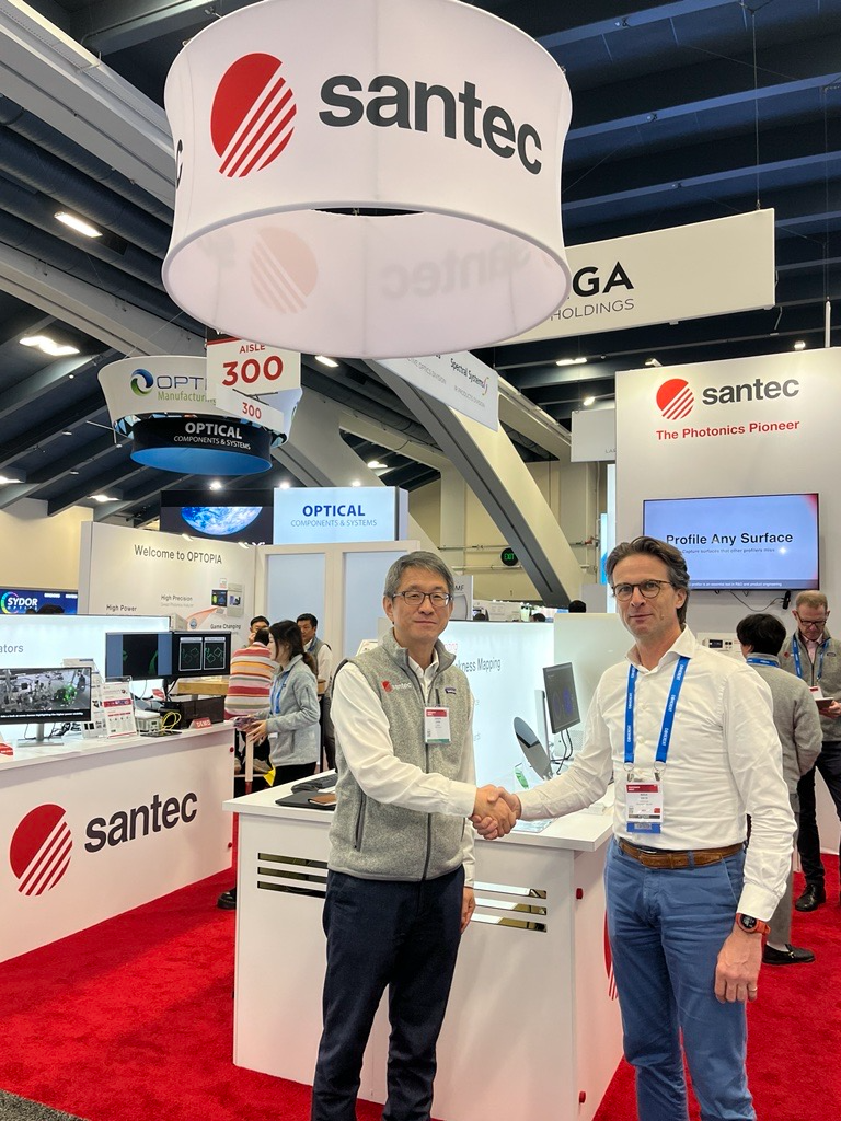
News
Contact us
We would be happy to tell you more about our products
Our location
Torenallee 20 5617 BC Eindhoven
Call us
+31 6 4609 3997
Mail us
info@scinvivo.com